

What Is A Radioisotope Apex
How are radioisotopes used?
Radioisotopes are an essential part of radiopharmaceuticals. In fact, they have been used routinely in medicine for more 30 years. Every Australian is likely to benefit from nuclear medicine and, on average, will accept at least two nuclear medicine procedures in their lifetime[one].
Some radioisotopes used in nuclear medicine have brusk one-half-lives, which ways they decay quickly and are suitable for diagnostic purposes; others with longer half-lives take more fourth dimension to decay, which makes them suitable for therapeutic purposes.
Industry uses radioisotopes in a diversity of means to improve productivity and gain data that cannot be obtained in any other way.
Radioisotopes are commonly used in industrial radiography, which uses a gamma source to behave stress testing or check the integrity of welds. A common example is to test aeroplane jet engine turbines for structural integrity.
Radioisotopes are also used past industry for gauging (to measure levels of liquid inside containers, for case) or to measure out the thickness of materials.
Radioisotopes are likewise widely used in scientific research and are employed in a range of applications, from tracing the flow of contaminants in biological systems to determining metabolic processes in small-scale Australian animals.
They are likewise used on behalf of international nuclear safeguards agencies to notice clandestine nuclear activities from the distinctive radioisotopes produced by weapons programs.
What is a radioactive source?
A sealed radioactive source is an encapsulated quantity of a radioisotope used to provide a beam of ionising radiations. Industrial sources normally contain radioisotopes that emit gamma rays or X-rays.
What are some commonly-used radioisotopes?
Radioisotopes are used in a variety of applications in medical, industrial, and scientific fields. Some radioisotopes normally-used in industry and science can be establish in the tables below. Medical radioisotopes are described in the next section.
Naturally-occurring radioisotopes in manufacture and science
| Radioisotope | Half-life | Use |
|---|---|---|
| Hydrogen-3 (tritium) | 12.32 years | Used to mensurate the age of 'young' groundwater up to xxx years old. |
| Carbon-fourteen | v,700 years | Used to measure the age of organic material upward to l,000 years one-time. |
| Chlorine-36 | 301,000 years | Used to measure out sources of chloride and the age of water up to 2 one thousand thousand years old. |
| Lead-210 | 22.2 years | Used to date layers of sand and soil laid down upwards to lxxx years ago. |
Artificially-produced radioisotopes in industry and science
| Radioisotope | One-half-life | Utilize |
|---|---|---|
| Hydrogen-3 (tritium) | 12.32 years | Used as a tracer in tritiated water to written report sewage and liquid wastes. |
| Chromium-51 | 27.7 days | Used to trace sand to report littoral erosion. |
| Manganese-54 | 312.12 days | Used to predict the behaviour of heavy metal components in effluents from mining waste material water. Produced in reactors. |
| Cobalt-60 | five.27 years | Used in gamma radiography, gauging, and commercial medical equipment sterilisation. Besides used to irradiate fruit fly larvae in order to contain and eradicate outbreaks, as an alternative to the utilise of toxic pesticides. Produced in reactors. |
| Zinc-65 | 243.66 days | Used to predict the behaviour of heavy metal components in effluents from mining waste water. Produced in cyclotrons. |
| Technetium-99m | 6.01 hours | Used to written report sewage and liquid waste movements. Produced in 'generators' from the disuse of molybdenum-99, which is in plow produced in reactors. |
| Caesium-137 | 30.08 years | Used as a radiotracer to identify sources of soil erosion and depositing, and besides used for thickness gauging. Produced in reactors. |
| Ytterbium-169 | 32.03 days | Used in gamma radiography. |
| Iridium-192 | 73.83 days | Used in gamma radiography. Besides used to trace sand to study littoral erosion. Produced in reactors. |
| Gold-198 | two.lxx days | Used to trace sand movement in river beds and on sea floors, and to trace sand to study littoral erosion. Likewise used to trace manufactory waste causing bounding main pollution, and to written report sewage and liquid waste movements. Produced in reactors. |
| Americium-241 | 432.5 years | Used in neutron gauging and fume detectors. Produced in reactors. |
Radioisotopes in medicine
Nuclear medicine uses small amounts of radiation to provide information about a person's torso and the functioning of specific organs, ongoing biological processes, or the disease state of a specific illness. In virtually cases the information is used by physicians to make an accurate diagnosis. In sure cases radiation can be used to treat diseased organs or tumours.
How are medical radioisotopes made?
Medical radioisotopes are made from materials bombarded by neutrons in a reactor, or by protons in an accelerator called a cyclotron. ANSTO uses both of these methods. Radioisotopes are an essential part of radiopharmaceuticals. Some hospitals have their ain cyclotrons, which are generally used to brand radiopharmaceuticals with brusk half-lives of seconds or minutes.
What are radiopharmaceuticals?
A radiopharmaceutical is a molecule that consists of a radioisotope tracer attached to a pharmaceutical. After entering the body, the radio-labelled pharmaceutical will accumulate in a specific organ or tumour tissue. The radioisotope attached to the targeting pharmaceutical will undergo decay and produce specific amounts of radiation that tin exist used to diagnose or care for human diseases and injuries. The amount of radiopharmaceutical administered is carefully selected to ensure the condom of each patient.
Common radiopharmaceuticals
About 25 different radiopharmaceuticals are routinely used in Australia's nuclear medicine centres.
The most common is technetium-99m, which has its origins as uranium silicide sealed in an aluminium strip and placed in the OPAL reactor's neutron-rich reflector vessel surrounding the cadre. Later on processing, the resulting molybdenum-99 precursor is removed and placed into devices called technetium generators, where the molybdenum-99 decays to technetium-99m. These generators are distributed by ANSTO to medical centres throughout Australia and the about Asia Pacific region.
A short half-life of vi hours, and the weak energy of the gamma ray it emits, makes technetium-99m ideal for imaging organs of the body for disease detection without delivering a significant radiation dose to the patient. The generator remains constructive for several days of employ and is then returned to ANSTO for replenishment.
Another radiopharmaceutical produced in OPAL is iodine-131. With a one-half-life of eight days, and a college-energy beta particle decay, iodine-131 is used to treat thyroid cancer. Because the thyroid gland produces the body's supply of iodine, the gland naturally accumulates iodine-131 injected into the patient. The radiations from iodine-131 then attacks nearby cancer cells with minimal effect on salubrious tissue.
Other commonly-used radiopharmaceuticals can be constitute in the lists beneath.
Reactor-produced medical radioisotopes
| Radioisotope | Half-life | Use |
|---|---|---|
| Phosphorus-32 | xiv.26 days | Used in the treatment of excess red blood cells. |
| Chromium-51 | 27.lxx days | Used to label red claret cells and quantify gastro-abdominal protein loss. |
| Yttrium-90 | 64 hours | Used for liver cancer therapy. |
| Molybdenum-99 | 65.94 hours | Used equally the 'parent' in a generator to produce technetium-99m, the almost widely used radioisotope in nuclear medicine. |
| Technetium-99m | vi.01 hours | Used to prototype the brain, thyroid, lungs, liver, spleen, kidney, gall bladder, skeleton, blood puddle, bone marrow, heart claret puddle, salivary and lacrimal glands, and to notice infection. |
| Iodine-131 | eight.03 days | Used to diagnose and treat various diseases associated with the human being thyroid. |
| Samarium-153 | 46.28 hours | Used to reduce the pain associated with bony metastases of primary tumours. |
| Lutetium-177 | 6.65 days | Currently in clinical trials. Used to treat a variety of cancers, including neuroendocrine tumours and prostate cancer. |
| Iridium-192 | 73.83 days | Supplied in wire form for utilize as an internal radiotherapy source for certain cancers, including those of the head and chest. |
Cyclotron-produced medical radioisotopes
| Radioisotope | Half-life | Use |
|---|---|---|
| Carbon-11 | xx.33 minutes | Used in Positron Emission Tomography (PET) scans to study brain physiology and pathology, to find the location of epileptic foci, and in dementia, psychiatry, and neuropharmacology studies. As well used to detect heart problems and diagnose certain types of cancer. |
| Nitrogen-13 | ix.97 minutes | Used in PET scans as a claret menstruation tracer and in cardiac studies. |
| Oxygen-15 | 2.04 minutes | Used in PET scans to characterization oxygen, carbon dioxide and water in order to measure out claret flow, blood volume, and oxygen consumption. |
| Fluorine-18 | 1.83 hours | The about widely-used PET radioisotope. Used in a variety of research and diagnostic applications, including the labelling of glucose (equally fluorodeoxyglucose) to notice encephalon tumours via increased glucose metabolism. |
| Copper-64 | 12.7 hours | Used to study genetic illness affecting copper metabolism, in PET scans, and also has potential therapeutic uses. |
| Gallium-67 | 78.28 hours | Used in imaging to detect tumours and infections. |
| Iodine-123 | thirteen.22 hours | Used in imaging to monitor thyroid function and detect adrenal dysfunction. |
| Thallium-201 | 73.01 hours | Used in imaging to detect the location of the damaged eye muscle. |
Nuclear imaging
Nuclear imaging is a diagnostic technique that uses radioisotopes that emit gamma rays from within the body.
How is nuclear imaging different to other imaging systems?
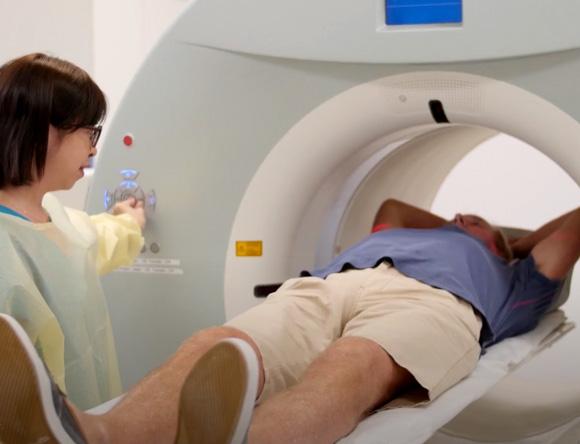
There is a pregnant deviation betwixt nuclear imaging and other medical imaging systems such as CT (Computed Tomography), MRI (Magnetic Resonance Imaging) or X-rays.
The main difference between nuclear imaging and other imaging systems is that, in nuclear imaging, the source of the emitted radiations is within the body. Nuclear imaging shows the position and concentration of the radioisotope. If very little of the radioisotope has been taken up a 'cold spot' volition prove on the screen indicating, perhaps, that blood is not getting through. A 'hot spot' on the other mitt may betoken excess radioactivity uptake in the tissue or organ that may exist due to a diseased land, such as an infection or cancer. Both bone and soft tissue can exist imaged successfully with this system.
How does nuclear imaging work?
A radiopharmaceutical is given orally, injected or inhaled, and is detected by a gamma camera which is used to create a computer-enhanced image that can be viewed by the physician.
Nuclear imaging measures the officeof a part of the body (by measuring blood flow, distribution or accumulation of the radioisotope), and does not provide highly-resolved anatomical images of body structures.
What can nuclear imaging tell us?
The information obtained by nuclear imaging tells an experienced dr. much about how a given office of a person's torso is performance. By using nuclear imaging to obtain a bone scan, for case, physicians tin detect the presence of secondary cancer 'spread' up to ii years ahead of a standard X-ray. It highlights the almost microscopic remodelling attempts of the skeleton as it fights the invading cancer cells.
Other types of imaging
Positron Emission Tomography (PET) scans
A widely-used nuclear imaging technique for detecting cancers and examining metabolic activeness in humans and animals. A small-scale corporeality of brusque-lived, positron-emitting radioactive isotope is injected into the body on a carrier molecule such as glucose. Glucose carries the positron emitter to areas of high metabolic action, such as a growing cancer. The positrons, which are emitted quickly, grade positronium with an electron from the bio-molecules in the trunk and then annihilate, producing a pair of gamma rays. Special detectors can runway this procedure, enabling the detection of cancers or abnormalities in brain function.
Computed Tomography (CT) scans
A CT scan, sometimes called CAT (Computerised Axial Tomography) browse, uses special X-ray equipment to obtain image information from hundreds of different angles around, and 'slices' through, the body. The information is and then processed to show a 3-D cross-section of torso tissues and organs. Since they provide views of the body piece by slice, CT scans provide much more comprehensive information than conventional X-rays. CT imaging is specially useful because it can show several types of tissue - lung, bone, soft tissue and claret vessels - with greater clarity than Ten-ray images.
Though a CT scan uses radiation, it is not a nuclear imaging technique, because the source of radiations - the Ten-rays - comes from equipment outside the body (as opposed to a radiopharmaceutical inside the body).
PET scans are frequently combined with CT scans, with the PET scan providing functional information (where the radioisotope has accumulated) and the CT scan refining the location. The primary advantage of PET imaging is that it tin provide the examining medico with quantified information about the radiopharmaceutical distribution in the absorbing tissue or organ.

What Is A Radioisotope Apex,
Source: https://www.ansto.gov.au/education/nuclear-facts/what-are-radioisotopes
Posted by: loftonbetwou.blogspot.com
0 Response to "What Is A Radioisotope Apex"
Post a Comment